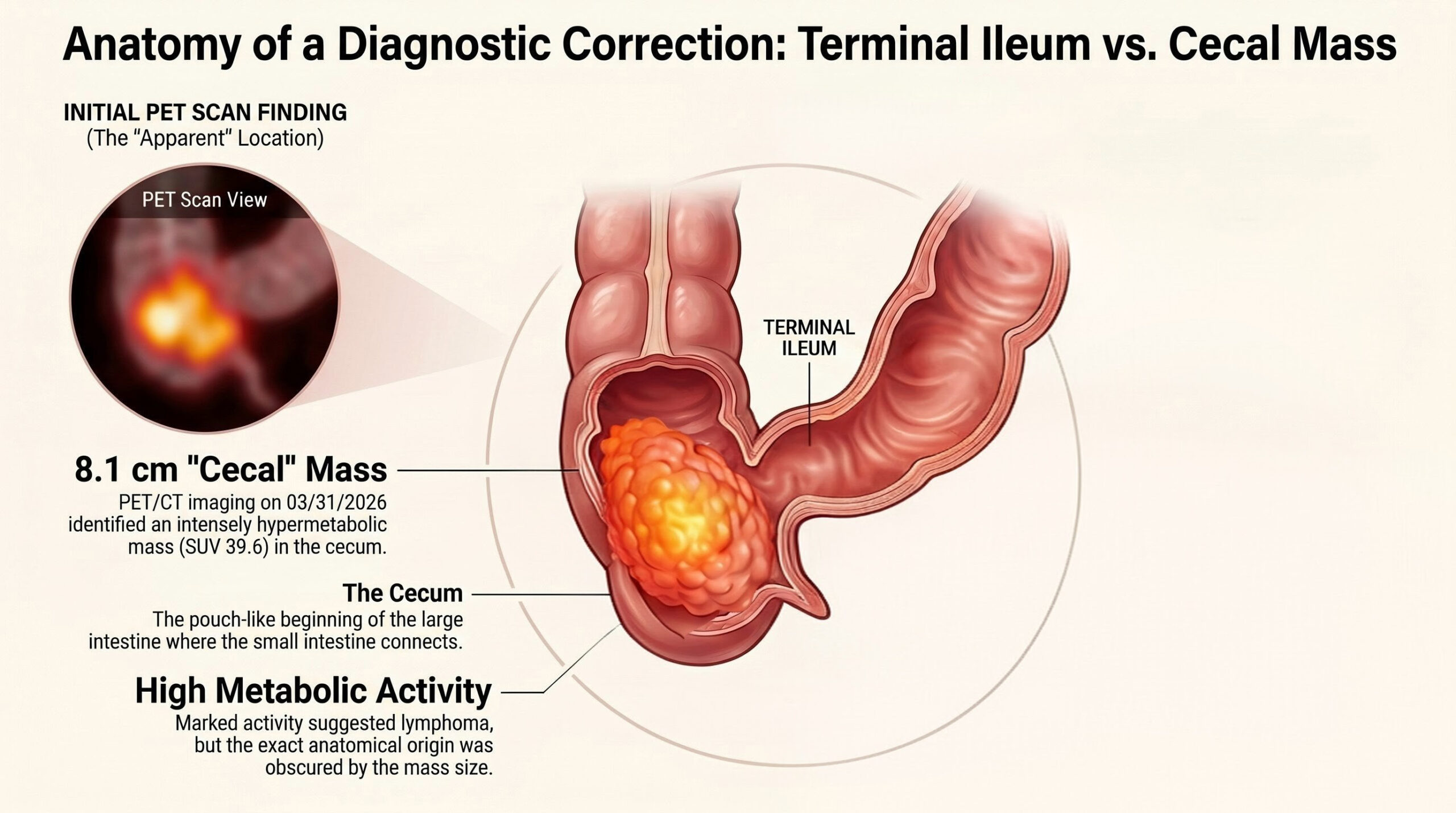
This is what we went into biopsy expecting them to find, based on the petscan.
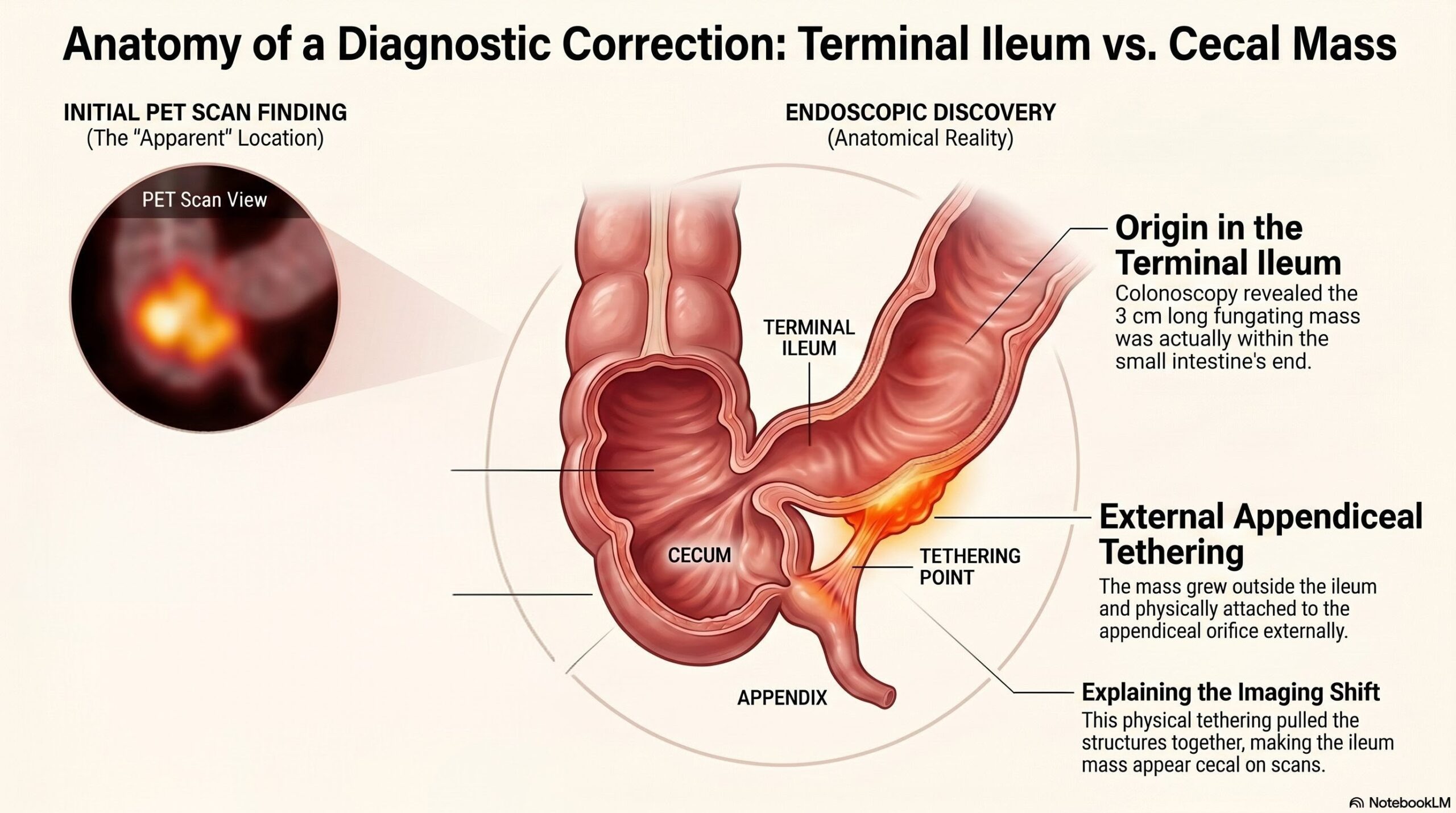
What the colonoscopy results state they found once there, with the cecum completely clear. The words spoken in recovery were clearly indicating that they saw a little tension in the wall but that it was not obstructed, it was pulled a little and the area was shiny or slimy or something, which they described as ‘a little seepage’. They suspected that it was a lymphoma that had reached out either to or from the appendix and bound them together, but it was only 3cm shiny spot that they saw, they saw nothing of the large 8.1cm grapefruit sized mass they predicted they would find.
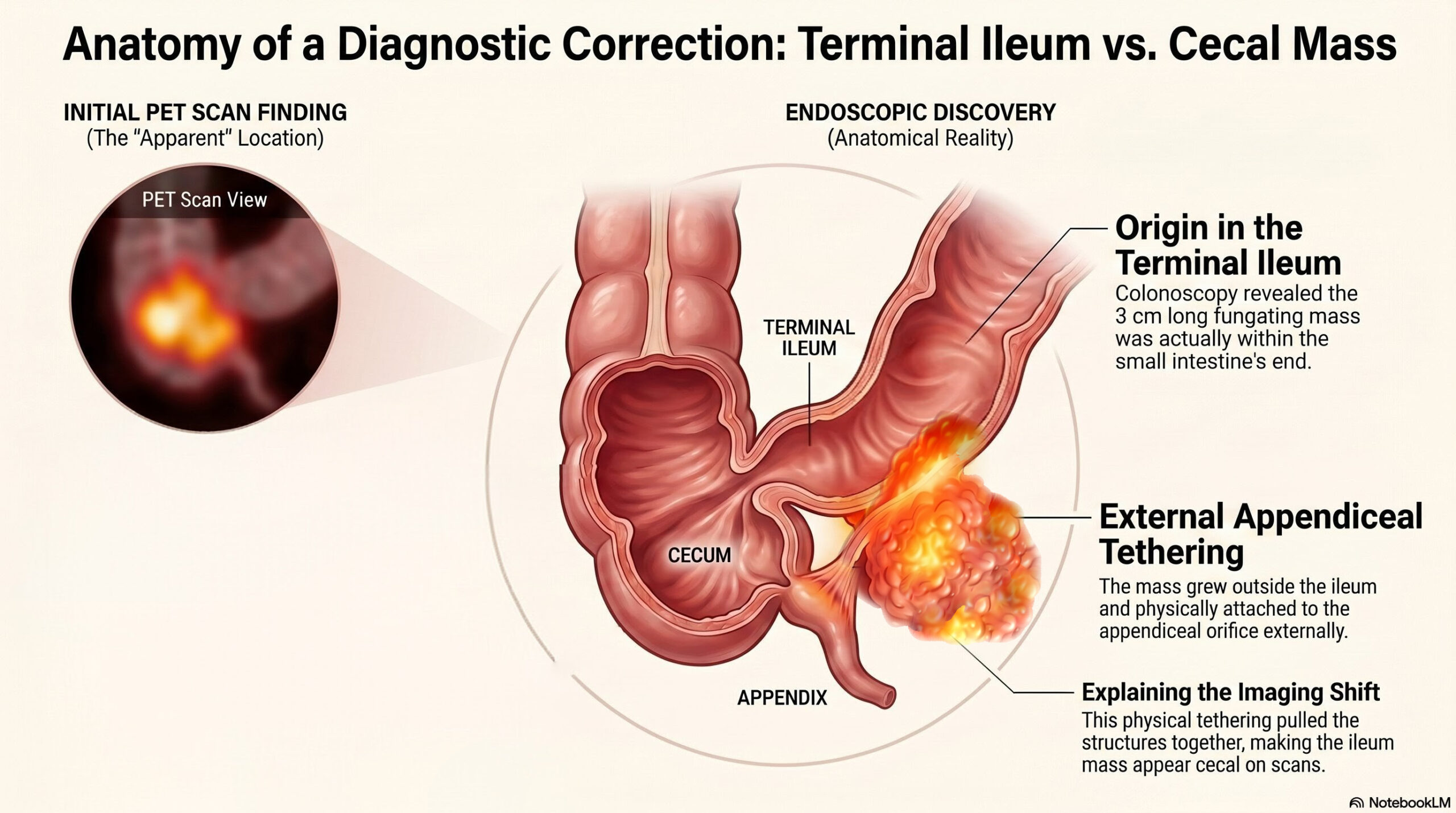
What I personally believe is actually there. They could not see outside the colon. They only saw a small 3cm shiny seepage spot on the wall of the Terminal Ileum. I figure that it could not easily make that huge bright spot on the imaging if it were not bigger outside the visible area, the iceberg effect, you only see what’s above the surface, you have no idea what is below. It could even be much smaller than their report. There’s no way to really know based on that camera they put through there. They did say they clipped several biopsy bits and that there was no blood at all involved in that or the rest of the procedure. That’s interesting, and probably noteworthy.
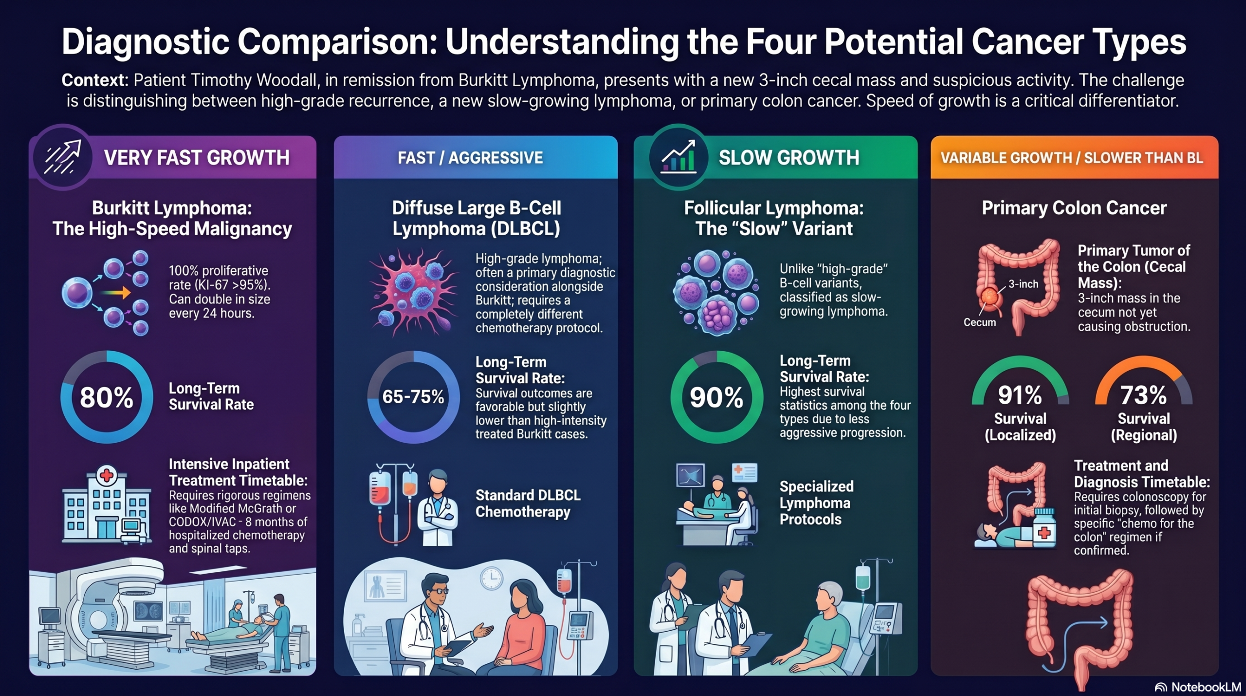
These are the what the biopsy will tell us when the results arrive, which one of these that it is. It’s unlikely that it’s berkitt’s. Previously, berkitt’s had been misdiagnosed as Follicular at Lenox Hill Hospital. That’s a possibility, and the best case scenario for treatment. Colon cancer is highly unlikely based on the location and the behavior. It could be the second one easily as well.
Doctors Dismissed Timothy’s Eight Centimeter Tumor